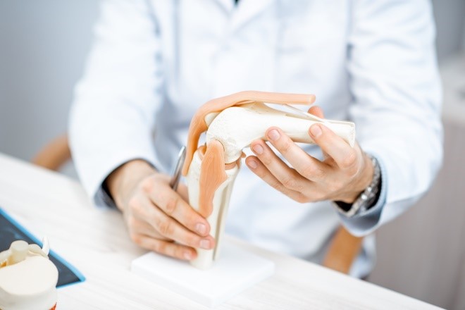
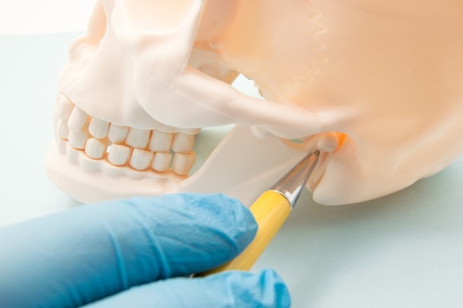
Joints like knees, shoulders, ankles, and the jaw connect our bones together into a functioning human body and allow us to move and bend. Over the course of life, joints are constantly in motion supporting every aspect of our functioning throughout the day. Injured, malformed, or worn-down joints create pain. Many people take opioid medications to provide relief from chronic joint pain.
The Helping to End Addiction Long-term® Initiative, or NIH HEAL Initiative,® is using several approaches to improve treatment for the millions of Americans who experience chronic pain, including that which affects joints. In particular, the Restoring Joint Health and Function to Reduce Pain (RE-JOIN) Consortium is using cutting-edge technology to expand our understanding of pain signaling in joints through a comprehensive characterization of sensory neurons (nerve cells) that allow us to feel pain. RE-JOIN scientists are focusing on two joints that often create chronic pain: the knee and the jaw, or temporomandibular (TMJ) joint.
- Which subtypes of nerves are distributed throughout the knee and jaw joints?
- What molecular properties distinguish healthy and damaged joints?
- How does nerve wiring reorganize in response to changes in joints?
HEAL scientists are using a variety of innovative techniques to unlock the answers to these questions, toward identifying non-opioid treatment strategies for chronic pain.

Mapping the Knee Joint
One group led by Brendan Lee, M.D., Ph.D., of the Baylor College of Medicine, is using a technique (in mice) that labels neurons and captures a 3D image of the entire leg and part of the spine. The scientists will compare information gathered from mice at different ages, mice with osteoarthritis, and mice that have been given treatment for pain relief. Through this work, the team hopes to understand how pain-sensing neurons are distributed throughout the different tissues of the mouse joint (or “innervation”), map their pathways and connection to the brain and spinal cord, and elucidate how the wiring reorganizes under different conditions.
Other research looking at the knee joint, led by Anne-Marie Malfait, M.D., Ph.D., of Rush University is mapping innervation of both mouse and human knee joints. This group is using state-of-the-art imaging along with transcriptomics, a technique that measures gene activity driving neural network reorganization. In addition to comparing tissues from healthy and diseased mice, they will also examine (from both younger individuals with no signs of osteoarthritis and from older individuals expected to have developed the condition at some point) donated tissue from the knee as well as from the dorsal root ganglia, clusters of neurons near the spinal cord.
Information gathered through this research will be used to build detailed 3D models of knee joint sensory networks, create a chart of neurons that innervate the knee, and document interactions between nerve and joint cells. The approach will help scientists understand precisely how joint innervation changes in injury and disease.
“Being able to manipulate these key elements should lead to completely novel therapies for improving joint health and reducing joint pain,” explains Malfait.

Mapping the TMJ (Jaw) Joint
HEAL researchers also aim to get a comprehensive picture of nerve wiring of the jaw, toward understanding how pain develops in this complex joint that helps control breathing, eating, and speech. Like with the knee joint, there is a lot to learn.
Christopher Donnelly, D.D.S., Ph.D., of Duke University leads a RE-JOIN research team that is using magnetic resonance imaging (MRI) to map the sensory neural networks of jaw tissues in both mice and humans. The scientists aim to find the sensory neurons innervating the TMJ in both healthy mice and those with jaw disorders associated with acute and chronic pain. They will also analyze human jaw samples to see how different types of neurons contribute to the development of pain at a molecular level.
Another RE-JOIN project led by Armen Akopian, Ph.D., of the University of Texas Health Sciences Center at San Antonio, is focusing on identifying the function, type, distribution, and molecular signatures of a group of specific nerves that provide sensation to the face and neck, including jaw tissues. One goal of this work, says Akopian, is to develop good animal models for TMJ disorders, including chronic pain – and ultimately reduce the need for opioid treatment of joint disorders.
Mapping the Disease–Pain Relationship
Kyle Allen, Ph.D., of the University of Florida, is on the case, using advanced behavioral testing of rodents to assess how changes in joint innervation affect behaviors like movement and motion, as well as how they interact with each other. The scientists will also measure joint function and conduct sensory tests in human patients with jaw pain to see how neural network rewiring affects an individual’s pain symptoms.
“This research will help us stratify patients and identify the most effective personalized treatments,” says Allen, “as well as potentially develop new therapies in general for chronic knee and TMJ pain.”
NIAMS
Learn about the role that the National Institute of Arthritis and Musculoskeletal and Skin Diseases (NIAMS) is playing in the NIH HEAL Initiative.
Sign Up to Receive HEAL Research News
Subscribe to the monthly HEAL Digest to receive NIH HEAL Initiative research advances in your inbox.
 U.S. Department of Health & Human Services
